


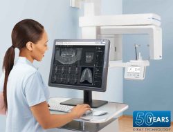
Description
See All That You Need To See
- Exceptional 3D & 2D X-ray image quality
- The anatomically adapted jaw-shaped 3D Field of View (FOV) captures the entire dentition, including third molar area – The Scope of Dentistry
- 50 x 50 mm volumes in 80 or 120 μm resolution – Great for Single Implants and Endo
- Highest resolution CsI (Cesium Iodide) enhanced sensor – creates brilliant, high-quality 3D & 2D images from one sensor, no swapping, no complications
Ideal Imaging FOVs | Easy Positioning | High Image Quality
- In addition to the standard adult 130mm x 85mm FOV, the ProVecta 3D Prime offers child size and 50mm x 50mm FOVs – great for Endo & Single Implants
- 80, 120,& 200 -microns resolution (pixel size) available to maximize resolution and clarity
- Reconstruction algorithms allow 3D volumes to be rendered and displayed in the shortest time possible to maximize clinical workflow
Maximize Your Diagnostic Capabilities
- Innovative 7” touchscreen provides clear text & symbols to guide you through all functions
- Open Architecture – Integration available with most for implant planning and surgical guide designing softwares (Exocad®, SimPlant®, plus more)
- Simplified Positioning thanks to double laser alignment for 3D scans & triple laser alignment for 2D scans
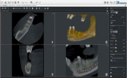
- Easily verify implant positioning, root, and jaw fractures diagnoisis, and more
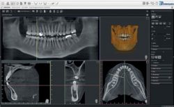
- VisionX 2D/3D software Included – a new state of the art and powerful tool
-
- Utilizes time saving Artificial Intelligence (AI)- Unique to VisionX
- Scan to Plan times shortest among competitors
- VisionX features three different 3D views (Panoramic, TSA, MPR)
- Accurate tracing of mandibular nerve canal (using AI – 30 second process)
- Easily measure anatomical structures with accuracy (no magnification)
- Import & export of competitor’s 3D DICOM datasets – Versatility
- No annual support fees
ProVecta 3D Prime covers the entire dentition all patients
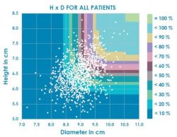
White Paper study results:
- 1,020 patients were examined in a study from Dr Johannes Krause*
- The study shows that a FOV with a height of 85 mm and diameter of 110mm is required for 100% coverage of the dentition (all 4 3rd molars – one exposure)
- With a competitively priced units with a FOV of 80 x 80 mm, only around 1.4% of all patients can the full dentition be captured in one scan (rarely are all four 3rd molars captured – usually two (2) scans are required)
By contrast, the adapted, jaw-shaped volume of the ProVecta 3D Prime covers the entire dentition in most patients – the Scope of Dentistry